
Beaumont Hospital, Grosse Pointe is employing a new two-step strategy to detect lung cancer using a radiation CT scan and Veran technology, which fuels electromagnetic navigational bronchoscopy, allowing doctors to take biopsies or mark abnormalities.
Believed to be the only such technology available in the region, the two-step process was introduced to the hospital in November as part of Lung Cancer Awareness month. The technology is designed to improve outcomes in a disease that doesn’t produce any symptoms early on and has a five-year survival rate of only 20 percent.
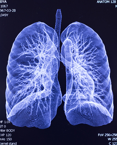
The first step of the procedure focuses on early detection through a low-dose radiation CT scan, which is available at multidisciplinary lung nodule clinics in Beaumont’s Royal Oak, Wayne, and Grosse Pointe hospitals. Lung nodules are lung abnormalities smaller than 3 centimeters in diameter. CT scans can detect nodules less than 1 centimeter in size. Longtime smokers, including those who have quit within the previous 15 years after smoking for a long time, qualify for an annual screening that is covered by insurance. Veran technology is the second step.
According to Dr. Michael Coello, medical director of the Lung Nodule Clinic at Beaumont, Grosse Pointe, surgeons can use the technology to pinpoint nodules and diagnose cancer in its earliest, most treatable stages.
“In the past, we used a digital photograph – a static snapshot – as our roadmap,” he says. “But we are moving, breathing beings, which means tumors also move with the person. As a result, precise tumor localization was difficult. This new technology keeps us locked in and synchronized with the patient’s breathing.”
The new technology also offers flexibility.
“If the nodule has no connection to an existing airway, in the past, we couldn’t access the lesion via bronchoscopy and would not even attempt a biopsy,” says Coello. “Now, based on this dynamic image, we can create our own pathway if none exists, and advance a needle through the chest wall directly into the lesion.”
At this point, a sample of the nodule can be obtained for diagnosis or markers can be placed around it so surgeons can find and remove it.
“It’s very satisfying to be able to identify lung nodules early through the screening process, and then, if necessary, follow up with bronchoscopy, thus speeding diagnosis and treatment,” says Coello. “This is what will finally make a difference in lung cancer survival.”
Coello says the technology also has potential diagnostic applications for vaping-related illness.
“If we aren’t exactly sure what’s going on, but vaping is suspected as the culprit, we could use the technology to sample lung tissue and identify the lipid droplets which are believed to be the cause of the diffuse lung injury, thus establishing cause of illness,” he says. “This process could solidify diagnosis, speeding and enhancing treatment.”
Beaumont has a total net revenue of $4.7 billion and consist of eight hospitals with 3,429 beds, 145 outpatient sites, nearly 5,000 physicians, 38,000 employees, and 3,500 volunteers.


